![]() 2024 HDR Scholarship: Curtin-Orthopaedic Research Foundation WA - Apply now
2024 HDR Scholarship: Curtin-Orthopaedic Research Foundation WA - Apply now
2018 Corrosion Photo Competition Awards
Art in Science
We believe that art is a humanizing force that enables us to push the frontiers of knowledge and foster innovation. Thus, in December 2018, we launched the first Corrosion Photo Competition Awards as part of the Chevron-Woodside Chair in Corrosion. The goal of the photo competition is to encourage students and researchers to share their achievements (and frustrations) with their peers, colleagues, friends, and the rest of the Curtin community.
Photos are judged not only by their perceived quality, composition, and technical merit but also by the reason that led the participants to select them. Each participant could enter two photos, and they were asked to explain why they picked them.
Below are the three winners of the 1st Academic Photo Competition. You can also see the recipients of the 2019 awards here.
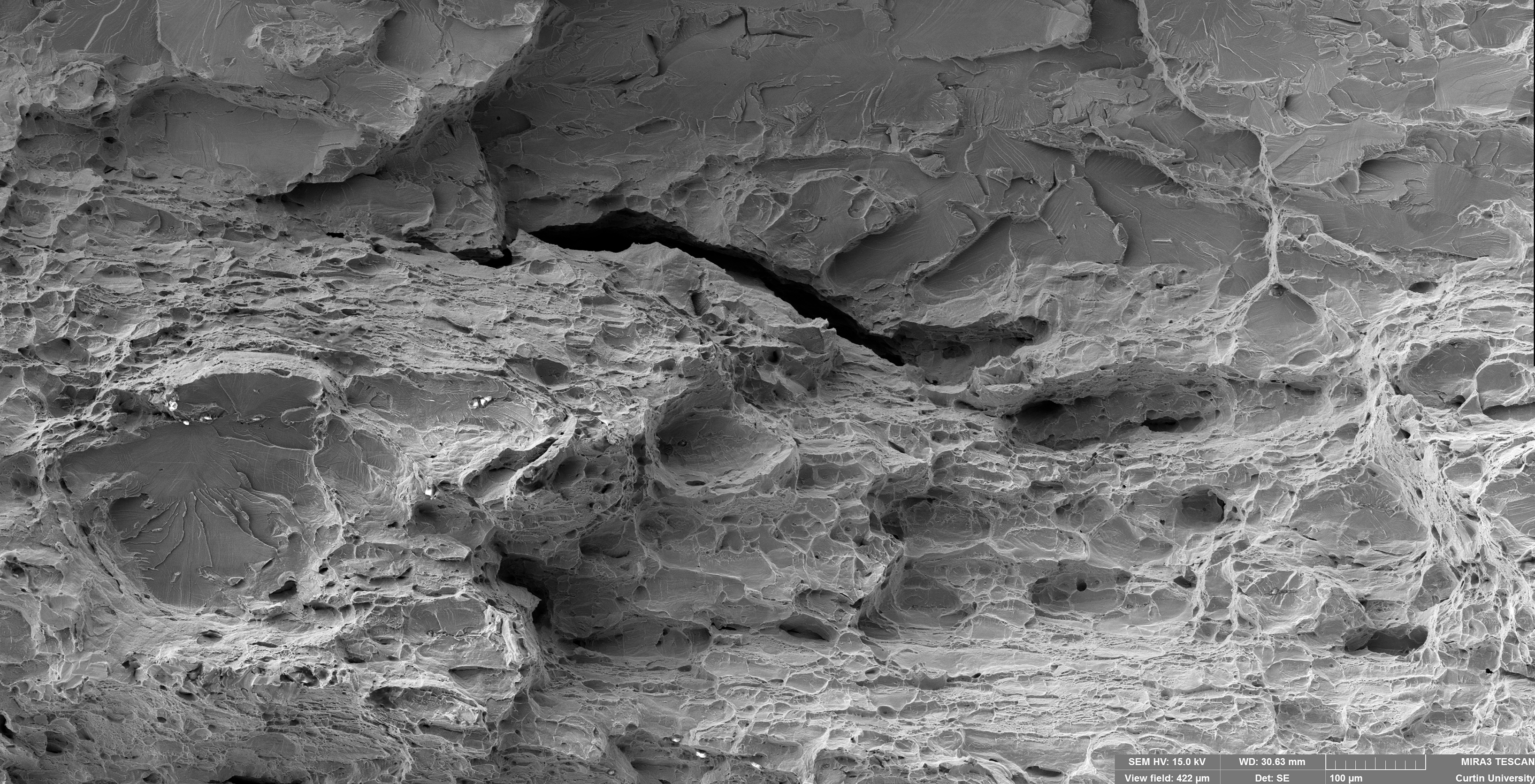
Third Place (a tie)
Edgar Hornus: The image is about the affected area by crevice corrosion in AISI 304L stainless steel, in chloride solutions at low temperature (0 °C). I used the PD-GS-PD (Potentiodynamic-Galvanostatic-Potentiodynamic) method with artificial crevice formers. This method is generally employed in nickel alloys or superalloys with high resistance to localized corrosion. In the image, we can see crevice corrosion with hexagonal pitting inside. Hexagonal pitting is probably due to the FCC crystalline structure of austenitic steels.
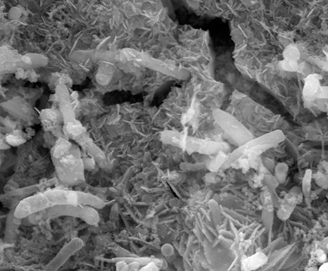
Hoda Ehsani: I like this photo because it shows how bacteria crosses through the crevice and caused pitting corrosion. The pit depth under the crevice was around 10 μm, and this picture shows that bacteria are responsible for it.
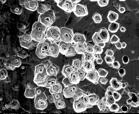
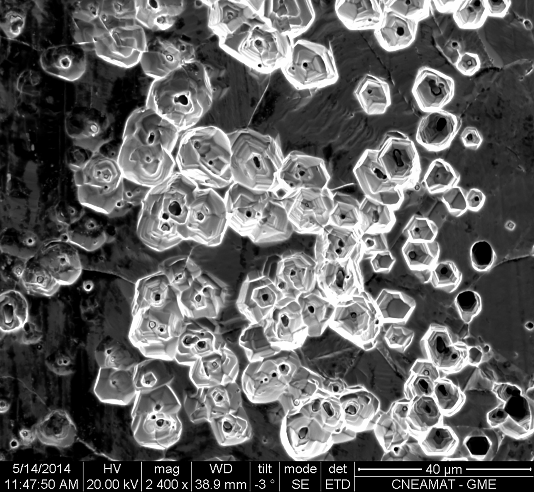
Third Place - Edgar Hornus: Secondary Electron SEM Image showing hexagonal pits on UNS S30403.
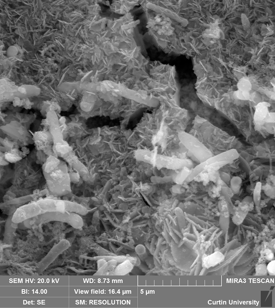
Third Place - Hoda Ehsani: FE-SEM image of microbially influenced corrosion formed on stainless steel (UNS S17400) crevice assembly samples exposed in artificial seawater in wet lay-up condition. The corrosion initiated by oxygen and was accelerated by bacteria.
Second place (a close call)
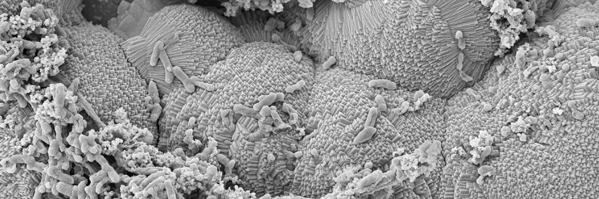
Erika Suarez: This bacterial isolate in certain conditions is able to extract electrons directly from the steel surface acceleration cathodic reaction, thus leading to electrical MIC (EMIC).
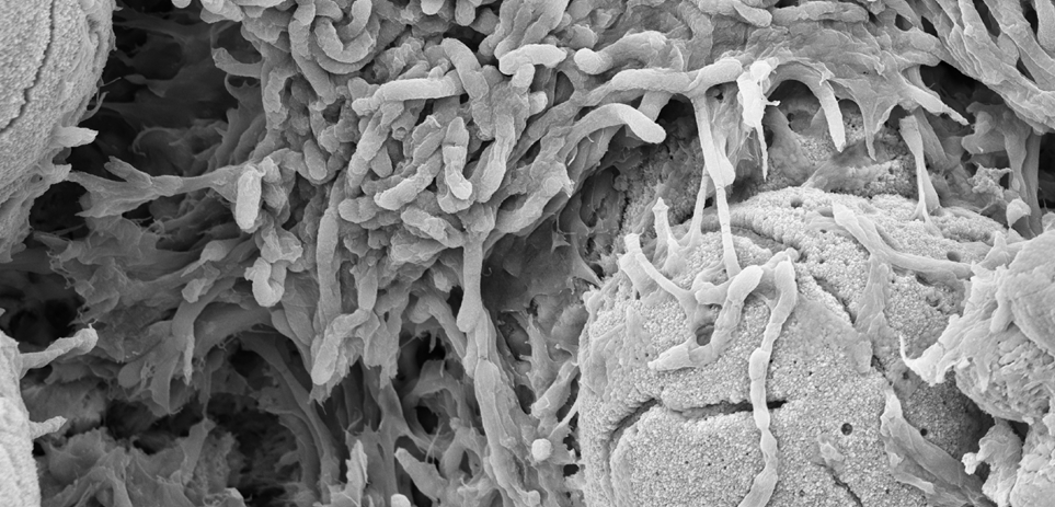
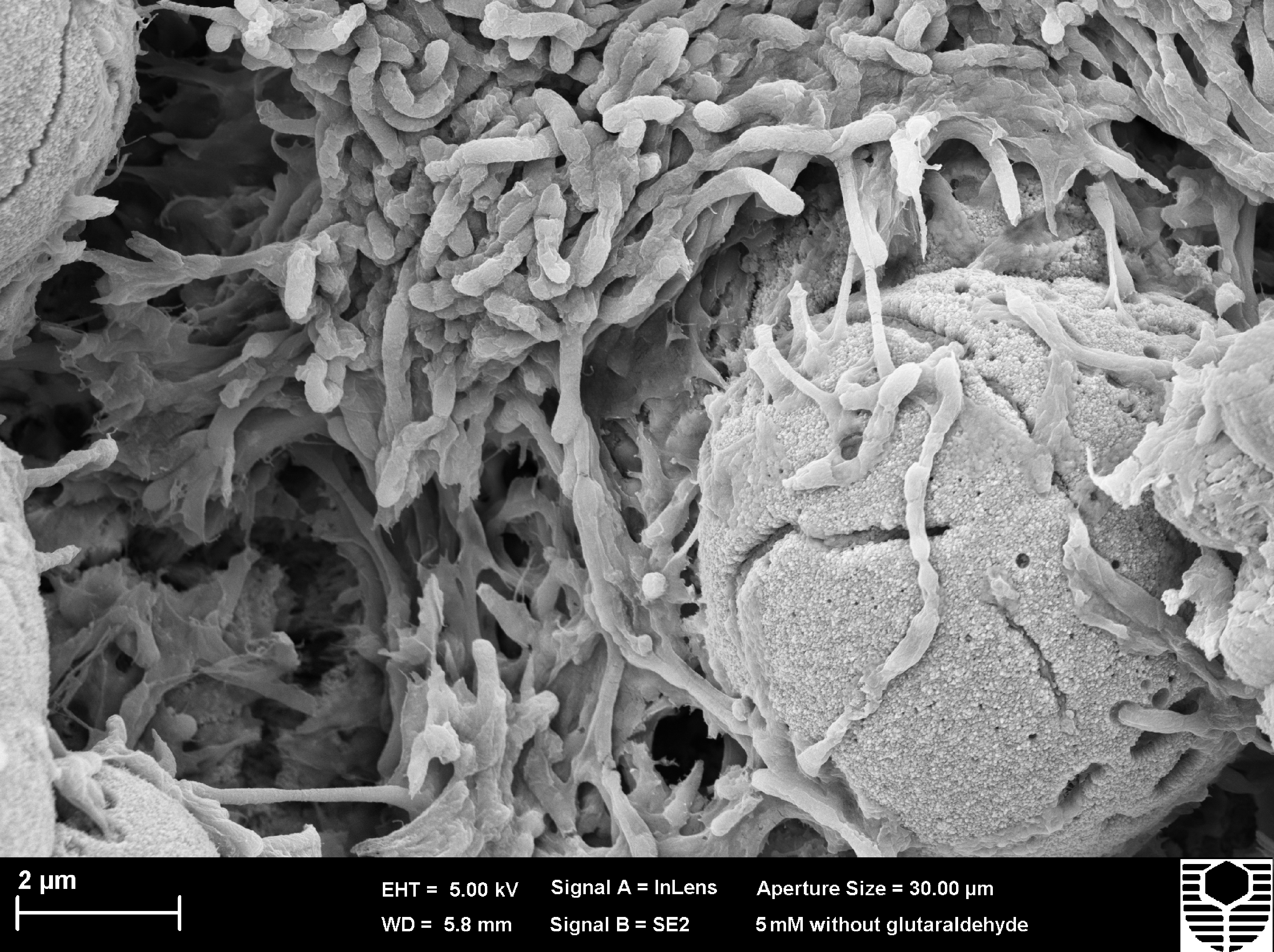
Second Place - Erika Suarez: FESEM image of biofilm formed (Shewanella oneidensis ATCC®700550) on carbon steel surface immerse in CO2/N2-saturated artificial seawater. Bacterial cells are surrounding iron oxides formed as a result of microbial activity.
First Place
María Sofía Hazarabedian: I have chosen this picture because it clearly shows the degradation of properties due to the hydrogen content as well as the effect of inclusions on metals. Finally but not less important, it represents personal progress for the sample preparation process and the acquisition of fractography images that are characterized to have a wide depth of field.
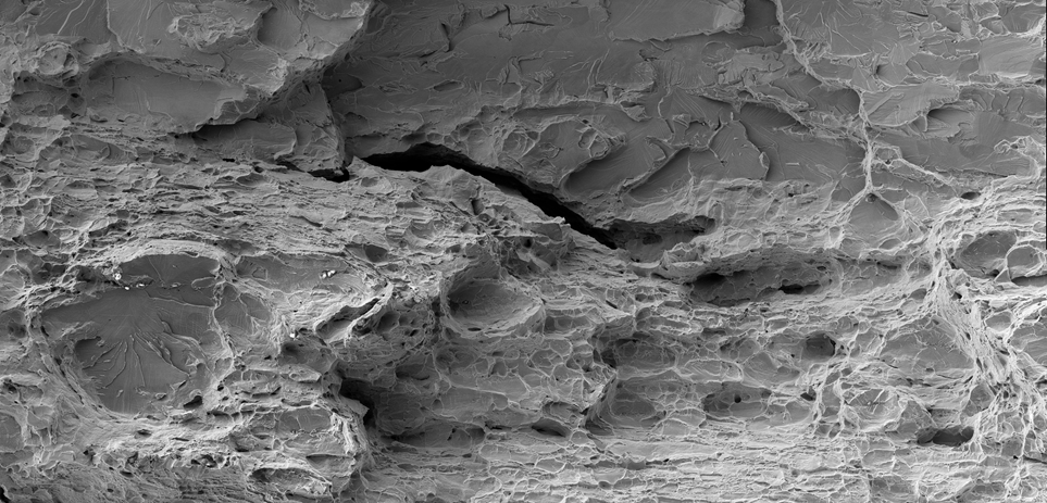
First Place - Sofía Hazarabedian: Composite secondary electron image of the fracture surface (full description in the following slides).
Acknowledgments
Many thanks to Prof. Claus Otto, Prof. Vladimir Golovanevskiy, Prof. Garry Leadbeater, Dr Zakaria Quadir, and Prof. Rolf Gubner for reviewing all the entries. Also, special thanks to Esteban Rodoni printing the photos for the exhibition, and for his photography, which we use across this website!